 Introduction
Determination of species of hypotrichs is a difficult task. The large number of species and their subtle differences in the arrangement of cirri complicate their correct determination. Helmut Berger, well known specialist for these species, already published four monographs of the hypotrichs. The collection of monographs is definetely a must have for the determination. The species Gastrostyla mystacea described here is listed in the first issue: "Monograph of the Oxytrichidae".
Discussion in the Austrian forum: Fluoreszenzfärbung von Gastrostyla mystacea
Determination using fluorescence staining
Gastrostyla mystacea belongs to the genus Oxytricha. They were found accompanied with another Oxtricha species of nearly the same size and shape from observation with the upright microscope. Such a mix of species in ciliate communities makes determination even more difficult.
This article shows the result of co-staining with Hoechst 33342 and acridine orange. This special staining method in combination with squash preparation and fluorescence excitation sometimes also yields staining of cirri of hypotrichs. Photomicrographys and wide field fluorescence microscopy will support exact determination of such species in these cases. The universal staining method is easy to use. Individuals can be observed using life-cell imaging over long periods of time to compare results in fluorescence and bright field imaging. Ongoing cell division can be observed as well.
Specific characters
Oxytrichs show patterns of cirri, that are typical for different species. The species have two marginal rows of cirri. These rows are found left and right to the ventral side of the cell. Front cirri, buccal cirri, caudal cirri and transversal cirri are found in groups. Further rows of cirri may appear in rows between the marginal rows.
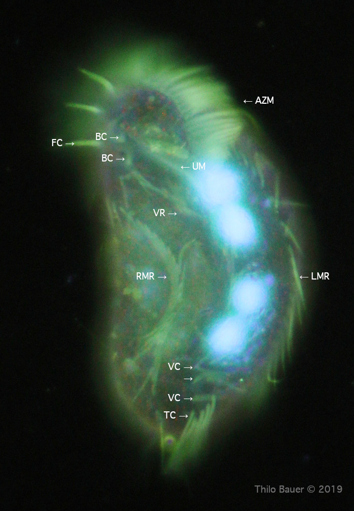
Gastrostyla has a prominent mid row of cirri starting short above the transversal cirri (5) up to the head of the cell. Typical for the species Gastrostyla mystacea are 3 separated ventral cirri close to the transversal cirri. Buccal cirri (2) are found at the top close to the front cirri (3). The peristome and adoral zone of membranes is also typical and froms a pattern like a question mark "?". See literature for further important details.
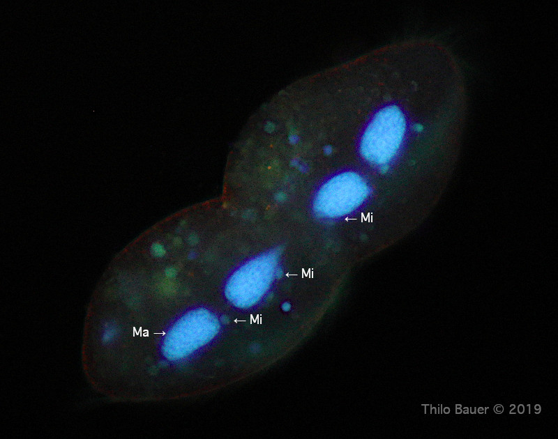
Gastrostyla mystacea shows two macronuclei (Ma), each accompanied by a micronucleus (Mi). The macronuclear anlage irritated me first, because images showed four macronuclei instead. Such a species must be determined as Gastrostyla steinii. However, the pattern of ventral cirri and transversal cirri and also the smaller size lead me finally to Gastrostyla mystacea. Another hint was given by the fact that many individuals were found in the stage of dividing cells. It is always impressive to see so many species dividing in fluorochromes that are told mutagene (breaking up DNA helix). From specific literature hints to DNA destruction from acridine orange are reported from investigations of isolated DNA. However, and from life-cell observations such cases are only described, if cells are irradiated and excited with bright, intensive ultraviolett light in addition. So the correct interpretation of the four macronuclei shown in my first picture is a cell division stage of this individual. Therefore the species correctly identifies as Gastrostyla mystacea.
My pictures seem to provide more than two micronuclei per indiviual cell. This kind of "artifact" probably can be explained as small phagosomes containing digested bacterial DNA. On the other hand, also micronuclei will divide, but at a different stage of the cell division and Mi may also temporarily disappear from observation. Such complications in addition make it even harder to determine the species. Such details about cell division should be known prior to species determination.
Legend
AZM: Adorale zone of membranelles
BC: Buccal cirri
FC: Frontal cirri
Ma: Macronucleus
Mi: Micronucleus
TC: Transversal cirri
VC: Pattern of three ventral cirri following transversal cirri
VR: Ventral middle row of cirri
Figure 1: Pattern of cirri in fluorescence co-staining with Ho342 and acridine orange. Four macronuclei may lead to wrong species determination (G. steinii), but is related to cell division.
Figure 2: Cell division stage observed in bright field illumination (oblique illumination). The middle ventral row of cirri (VR) can be well identified. The daughter cells already have formed two peristomes (AZM). This is the typical appearance of a cell division of hypotrichs.
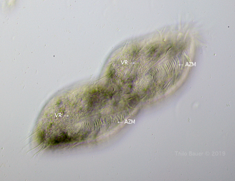
Bild 3: Same individual seen from fluorescence staining (UV excitation, 385 nm). Macronuclei in interphase appear more homogenuos and "with some holes". During cell division stage DNA will show in a chromosomal appearance, like seen in this fluorescence image. The outer border around the cell indicates small acidic granules below the pellicle.
Imaging details
Zeiss Axio Lab.A1, multi-immersions objektive Zeiss 40x/0,9 Ph3. Brightfield imaging with oblique illumination. Fluorescence: Triple-band filter cube F66-412, UV excitation at 385 nm. Camera: Canon EOS 77D.
Literature
- Berger, H. 1999, Monograph of the Oxytrichidae, Kluwer Academic Publishers.
- Foissner, W. et al. 1991. Taxonomische und ökologische Revision der Ciliaten des Saprobiensystems, Informationsberichte des Bayer. Landesamts f. Wasserwirtschaft.
- Bauer T. Fluoreszenz-Doppelfärbung mit Hoechst 33342 und Acridinorange zur Bestimmung der Arten von Ciliaten (Phylum: Ciliophora). Mikroskopie. 2019; 1: 2-19.