 Introduction
This article describes infection of Paramecium caudatum with bacteria of genus Holospora.
Description
Paramecium is the the well-known "slipper animalcule". Today, there exist at least 14 confirmed species, some of them considered as a complex of species. Paramecium is also one of only a few unicellular species, with well documented endoparasites or symbionts. Currently, about 60 different species are known to cause infections of Paramecium.
As a child doing some microscopy I ever was hoping to find Paramecium. It took me about 40 years to find the first Paramecia in one of my aquariums. Over years I found more and more sites with different Paramecium species, mainly P. caudatum, P. aurelia, P. bursaria and P. dubosqui. Reading books and articles about Paramecium, I was hoping to also find at least a single Paramecium infected by bacteria of genus Holospora. At the microscopy workshop in Inzigkofen this year (2020), I collected a sample from the river Donau. From this collected sample I observed the first Paramecium infected with Holospora.
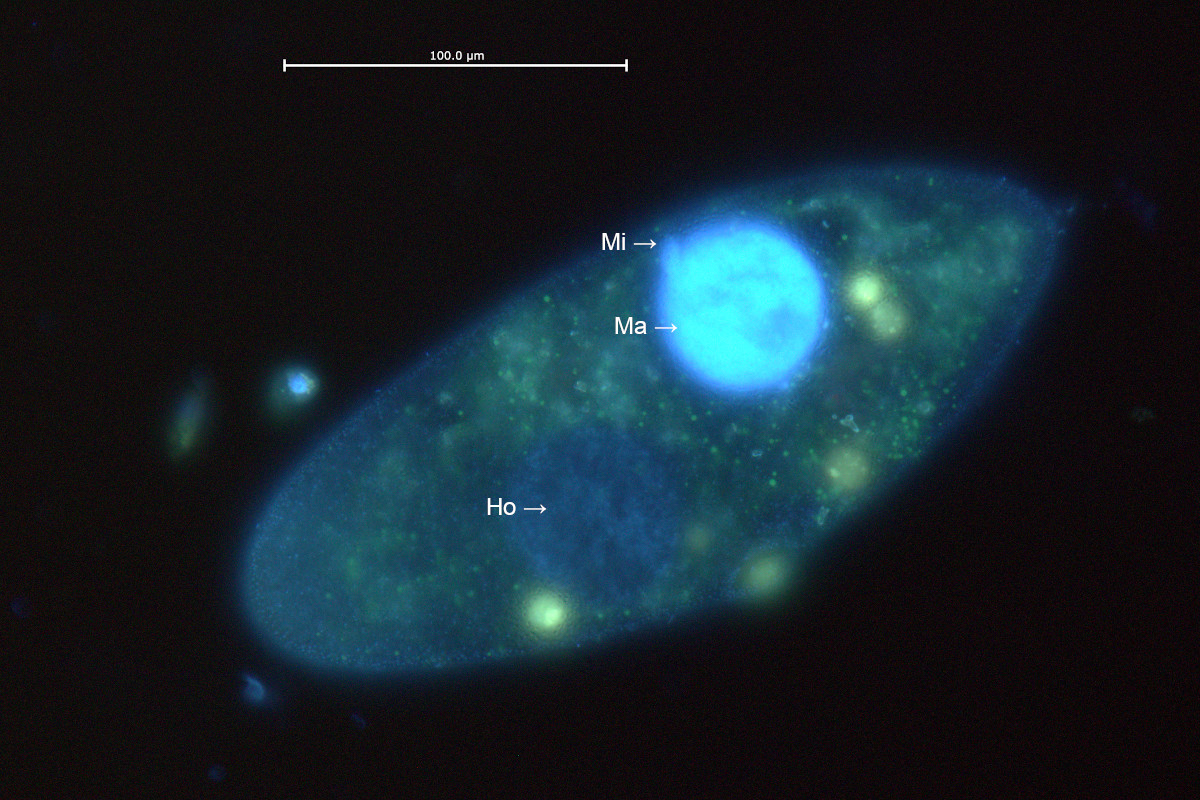
The single Paramecium individual shown here, demonstrates the way how this infection is spread between different individuals. Most ciliates species, also Paramecium, show nuclear dimorphism. This means ciliates have two different kinds of cellular nuclei, one or more macronuclei and one or more micronuclei. The two different types of nuclei of different size carry genetic material, but the purpose is different. Paramecium caudatum has a single macronucleus and a single micronucleus. Typically Paramecium is unicellular and proliferates by cell division. There also exists sexual reproduction by copulation of two individuals from time to time. In this case, genetic exchange and following fragmentation of the macronucleus will occur. A few consecutive cell divisions must occur, until the fragmented macronucleus will again merge into a single macronucleus. The individual shown here still shows two fragments of the macronucleus, which means it will take one more cell division until the nucleus merges back into a single one. It is interesting to see, only one of the two fragments carries bacteria of genus Holospora. This indicates the infection should be caused by an earlier copulation between a sane and an infected cell.
Legend:
- Ma: Macronucleus
- Mi: Micronucleus
- Ho: Fragment of macronucleus carrying Holospora
Figure 1: Bright field observation shows a "normal" fragment of the macronucleus (Ma, right) and a secondary fragment (Ho, left) infected with Holospora.

Figure 2:Detail of the macronucleus fragment infected.
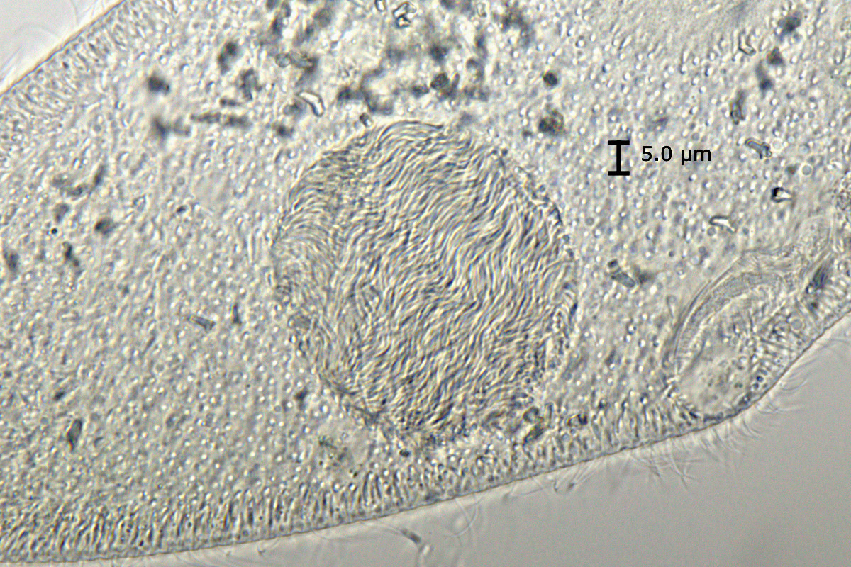
Figure 3: From fluorescence staining DNA of the normal macronucleus fragments is stained bright, while the fragment infected by Holospora shows a weak signal from staining of DNA and bacteria.
Photographic details
Zeiss AxioLab.A1, water immersion objektive C-Apochromat 40x/1,2 W Korr, Canon EOS 77D, fluorescence double staining using Hoechst 33342 and acridine orange.
Literature
- Fokin, S. I., & Görtz, H. D. (2009). Diversity of Holospora bacteria in Paramecium and their characterization. In Endosymbionts in Paramecium (pp. 161-199). Springer, Berlin, Heidelberg.
- Fokin, S. I. (2010). Paramecium genus: biodiversity, some morphological features and the key to the main morphospecies discrimination. Protistology, 6(4), 227-235.