 Introduction
Spirostomum species can be divided into two different sizes: Those species above 1 mm and others having half the size. Spirostomum belongs to the family of Heterotichidae.
Dicuss with us (in german): Austrian Tümplerforum.
Determination using fluorescence staining
Spirostomum minus has half the size of the similar species Sp. ambiguum, and measures a maximum of 600-700 µm. Size is an important character in this case. Spirostomum minus is claimed to be a complex of species, comparable to the Paramecium aurelia complex (Sonneborn). Reason to declare a complex of species is a certain variability within the group and similar, but not identical genetic properties, as discussed in literature (e.g. Foissner et al., 1992). Unfortunately, latest articles claim to have found similarities in genetic analysis. However, authors didn't demonstrate morphological features. Therefore, it is in doubt, if genetic classification is a valid character in this case, or if similar morphological features yield the status of a well defined, single genus. Genetic similarities are claimed based on small subunits of DNA, mostly 18S rDNA subunit (~3000 base pairs). On the other hand, the total content of DNA in ciliates is found to be in the order of 100 billions of base pairs (Prescott, 1994). These DNA subunits, however, show certain state of evolutionary development of individual species, which seems more reasonable.
I will show few images of a Sp. minus strain, that I recently collected in a pond of Hürth in the region between Bonn and Cologne. From a morphological description it certainly belongs to the Sp. minus clade.
This discovery is remarkably, as there was only a single cell of Sp. minus collected swimming between many Sp. teres, which could be taken into culture medium. Hoping it will grow, it showed amazing living will and grew up in simple culture medium to a rich colony of thousands of individuals in an erlenmayer flask within a week. In contrast to Sp. teres, Sp. minus has a moniliform macronucleus. So it could be well distinguished and isolated from the many Sp. teres under microscope at low magnification.
A detailed description of Spirostomum species is found with Boscaro et al. (2014). My German translation and remarks can be found here: Artenschlüssel von Spirostomum.
Figure 1: The length of the peristome of Sp. minus is about half the full length of the cell. It has a moniliform macronucleus, that is slightly visible in bright field. Adoral zone of membranelles and oral cilia can be seen up to the opening of the peristom (middle of image). Photograph taken in optimal oblique illumination.
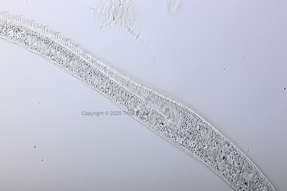
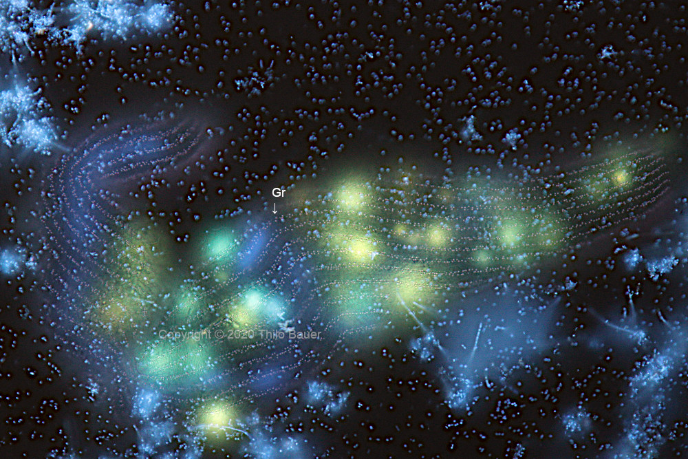
Figure 2: Fluorescent staining to demonstrate the moniliform macronucleus (Ma), many micronuclei (Mi) and phagosomes staing in green color (Ph). At the border of the cell stained granules (Gr) under pellicle is visible. See also description of figure 3.
Figure 3: Same individual as shown in figure 2. Microscope, however, is focussed to pellicle in this case. Granules are arranged in rows of 2-3 lines. Granules below pellicle appear acidic, because they are stained in red color by acridine orange. Below the pellicular granules other larger granules are visible, which appear not stained and statistically distributed. Looking closer to both fluorescence images, the larger granules, that are not stained, show a lens-like effect and small ring-shaped shadows against the unsharp organells found below these granules.
Imaging data
Photomicrographs taken with Zeiss Axio Lab.A1 FL-LED fluorescence microscope. Objektive Zeiss C-Apochromat 40x/1,2 W and condensor 1,2 were used for water immersion. Bright field image: Optimal oblique illumination. Fluorescence images: Double-staining with Ho342 and acridine orange, triple-band filter set F66-412, UV excitation (385 nm). Kamera: Canon EOS 77D.
Literatur
Boscaro, V., Carducci, et al., 2014. Focusing on genera to improve species identification: revised systematics of the ciliate Spirostomum. Protist, 165(4), 527-541.
Foissner, W. et al., 1992. Taxonomische und ökologische Revision der Ciliaten des Saprobiensystems, Informationsberichte des Bayer. Landesamts f. Wasserwirtschaft.
Prescott, D. M., 1994. The DNA of ciliated protozoa. Microbiol. Mol. Biol. Rev., 58(2), 233-267.