 Introduction
Microscopic observations of Spirostomum sp. show different small organelles below cuticle, especially between the ciliary rows. Staining with neutral red shows cortical organelles (granules) are acidic.
Experiment
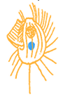
Neutral red is used in microscopy to demonstrate acidic pH in cell compartments. The dye is used at concentration 0,005% (1:20.000). Figure 1 shows staining of Spirostomum sp. culture no. 0016. Cortical granules are specific to determine certain Spirostomum species. (Foissner et al. 1992). It is in question, whether granules are identical with mucocysts. After staining many of the granules are missing between ciliary rows and large gaps appear.
Like many other supra-vital dyes, neutral red is toxic for ciliates. In acidic organelles the dye will stain organelles with red color. Spirostomum sp. suffer from intense microscopic illumination and will die soon, if stained with neutral red at given concentration. This is not the case, if individuals of that same species are observed without staining or using fluorescence dyes. Neutral red also shows fluorescence. Fluorescence of neutral red is quenched at larger concentration in acidic compartments: No fluorescence will be found, where organelles are showing red to dark red colors. The cytoplasm, however, shows bright fluorescence, which is no autofluorescence of the cell. Darker colors indicate larger concentrations of the dye (ion trap mechanism). It must be concluded that the dye is its own quencher at large concentration. It may also be concluded, that fluorescence of the dye and corresponding energy transfer in the cytoplasm is destructive and responsible for cell death. In contrast, typical fluorescence dyes are applied at much lower concentrations. This is the reason, why fluorescence dyes are less toxic.
Fig. 1: Spirostomum sp., stained with neutral red: Cortical granules are stained in red, carmine red to deep red colors. These organelles are acidic at pH<6,8. The dye probably induces ejection of mucocysts. This explains why there are large gaps in granular pattern between ciliary rows compared to bright field observations without staining or using fluorescence dyes.
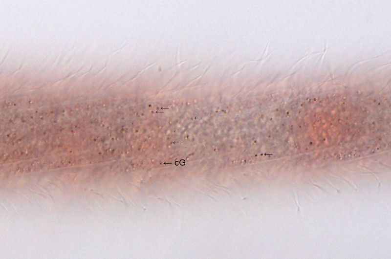
Fig. 2: Invisible death: Fluorescence of neutral red in cell compartments. Stained, acidic organelles do not show specific fluorescence, rather appear dark. This individual of Spirostomum sp. died quickly from fluorescence illumination. Energy transfer from fluorescence of the dye at high concentration micht be responsible for cell death and quick cell lysis. For comparison a bright field image of the same individual is shown.
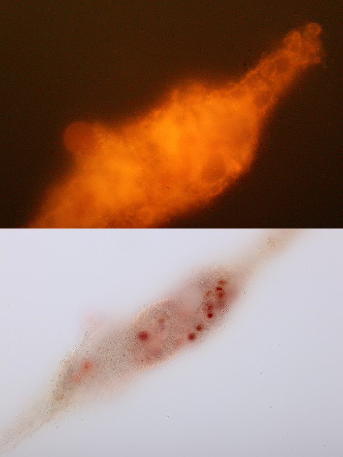
Fig. 3: Cortical granules (Gr) of Spirostomum sp. will also be stained by acridine orange in colors from green to red (UV excitation). This indicated, that acridine orange stains acidic cell organelles, rather that the nucleus, which is contrasted by another fluorescence dye: Hoechst 33342.
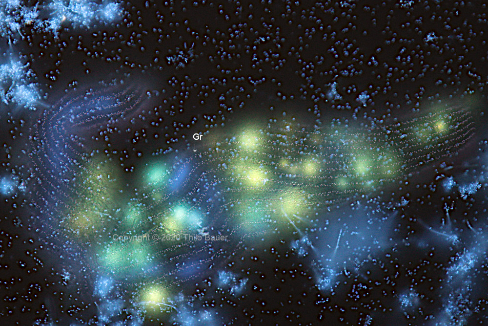
Fig. 4: Spirostomum sp.: fuorescence staining with acridine orange (blue excitation).
