
 Summary
The article describes methods to overcome spherical aberration in microscopy of water samples.
Introduction
Microscopists looking at planktonic life in water samples will most probably know this effect: Sometimes visual observation or photomicrography don't yield good microscopic images. Images look blurry and without contrast. With growing experience the observer notices: It depends on the height of the water column between the objective and the target. Over years, I always joked, my microscope is moody and has a bad day.
The plankton observer probably dealing with larger sample volumes starts to look through thick water columns from the beginning (typ. ≥100 µm). Sometimes this is caused by taking too large water drops or even the animals are relatively large, like large ciliates, small crabs or desmids. Certain observing techniques just require the use of larger volumes of water samples, like the micro-aquarium proposed by Michael Müller.
Link: Microaquarium proposed by Michael Müller (german only)
Under these circumstances standard microscope objectives do work for objects located close to the cover slide. The deeper the focal plane is located in the sample, the worse the image will look like. Classic microscope objectives are dry objectives or oil immersion objectives. While medium magnification (20x and below) will work passably well, the effect of blurred images starts to gain attention at a magnification of 40x. The effect will start to frustrate the observer while knowing, the objective is promised and designed to provide a high quality color correction and plane image, like a "fluar", "neo-fluar" or apochromatic objective. At deep water columns such objectives quickly will reach their limit. Colors, contrast and sharpness of the image drop dramatically. The focal plane feels undefined, important details no longer are seen. Now the observer might ask himself, if the objective is of poor quality.
Physical background
In the line of sight, light passes surfaces of glass, air, eventually immersion media and water (of the sample) and rough pellicles of the observed organisms. At every surface, between the different media light is refracted. The optical error that occurs here is named spherical aberration. Because of different refractive indices of media light beams passing the objective close to its border of the aperture pupil, near the optical axis and between the two extremes will form different focal planes (see image below).
Optical resolution of a microscope objective depends on a physical property called numerical aperture, called NA hereafter. The value of NA is printed onto the objective right after its magnification, e.g. "40x/0.60". Dry objectives have a practical limit at NA=0.95 (max.). Oil immersion objectives usually start from NA~1,0, up to a maximum NA=1,4. There are exceptions from the rule with extraordinary immersion objectives having large NA but low magnification, like the Zeiss life-cell imaging objectives LD LCI Plan-Apochromate 20x/0,8 (see also last section about water immersion objectives). Because of the larger NA, immersion objectives provide larger resolution, compared to dry objectives with similar magnification. That's why immersion objectives show more details. Immersion optics with NA~1.5 were already known in year 1892 (see: S. Bradbury, Evolution of the Microscope. 2014). Any NA larger than 1,4 is usually a special use case, like super-resolution imaging. They require special immersion media for both the immersion and medium of the sample. Hence, they do not work with simple cells in water (see below).
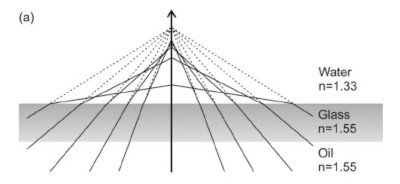
Figure 1: How spherical aberration occurs in water using an oil immersion objective. Because of different diffractive indices of the water medium different light rays result in different focal planes. Image taken from: Vermeulen, K. et al. 2006.
The optical error is called spherical aberration. The reason for an optical error like this is not the objective itself. The true reason is the behavior of light and the diffractive index of the water medium. Especially objectives with well corrected color aberration and plane focal image yield a good image only in a thin layer short below the cover slide of a microscopic sample. At deeper water columns, light gets more refracted in a different way. Thus, standard objectives provide a poor image quality at deeper water columns. Oil immersion objectives at large magnification are the most sensitive optics under such conditions because one expects them to provide finer details. However, when looking closer to details, like using digital image zoom of a digital photo, the error increases to its extreme.
Spherical aberration is an optical error, which is not caused by the objective, but the behavior of light which passes media with different refractive indices (see above illustration). Standard objectives do not compensate spherical aberration taking into account the embedding media of the sample. They work well only, if the immersion medium has the same refractive index as the sample medium. That said, standard oil immersion objectives work well only, if talking about durable preparations in resins like canada balsam or Euparal. Another use case covered by oil immersion are highly refractive sample media (see below). Oil immersion objectives simply are not designed for water medium. Used with water medium it is like with dry objectives: They provide good image quality only within a thin layer below the cover slide. With increasing depth in water spherical aberration increases quickly. Expecting good performance, the resulting image provides the opposite, when looking close onto fine details through thick water columns. Observed effects of spherical aberration are degraded contrast, increasing color aberration and loss of definition of the focal plane.
Strategies to avoid spherical aberration
So far we studied the effect and described its reason. From that countermeasures can be developed. From no-cost up to certain investment these are topics to consider:
- Reduction of numerical aperture
- Squash preparation
- Thin cover slides
- Precise sample volumes
- Highly refractive media
- The inverse microscope
- Water immersion objectives
They are described in the following sections.
Reduction of numerical aperture
In the older literature of protist imaging a method is described: Close the aperture of the condenser to improve contrast. This method comes at no cost. Indeed, reduction of the aperture of the condenser, and thus the aperture of the objective, seems to improves visual contrast. The effect is based on the fact that light rays at the border of the full aperture of the objective will be excluded. Thus, spherical aberration caused by light rays in the outer region of the aperture do not contribute any longer. As spherical aberration increases with the height of the water column, closing the aperture will not remove spherical aberration, but decrease optical resolution. Main disadvantage of this technique will be loss of details in the optical image. The objectives will not provide their full power in this case. Therefore, sorry to say it, this is not a method of choice. For photomicrography reduction of the numerical aperture is not recommended.
Squash preparation
If organisms are robust enough, squash preparation is proposed to obtain better results in photomicrography. The effect is based on the fact, that standard objectives are not designed for deep water columns, but work well in a small layer below the cover slide (see above). Therefore, squash preparation helps to circumvent spherical aberration by pushing the cover slide close enough to the target. With dry objectives evaporation of water will draw the cover slide to lower height. With oil immersion objectives the situation is more compley, however. The cover slide will be moved between the objective and target. With oil-immersion the exact location of the cover slide depends on the amount of immersion medium used and back pressure from the water sample. The effect can be seen as moving objects while focussing. This is not very well controllable. Hence, image quality is not well reproducible using oil immersion objectives. Serious comparison of different dry objectives or oil immersion objectives under these circumstances is simply impossible. Other disadvantages are: Many species are not robust enough and may collapse while trying to squash. Cells, once squashed, loose their natural shape. Testacea, desmids or small crabs are hard and cannot be squashed, or they just break.
Thin cover slides
Standard microscope objectives are designed to work with a thickness of cover slide of 0.17 mm. The cheaper cover slides offered in stores usually are thinner (0.13-0.16 mm). Because the goal is to reduce spherical aberration, thin cover slides may help to improve spherical aberration a bit. While standard slides are preferred (0.17 mm), the effect gained my attraction when looking into microscopes of colleagues, who use cheap glass slides for the observation of planktonic life. Often I got the impression the image look a bit more contrasted. Main disadvantages of this technique: Although not visible from the beginning, spherical aberration is still present. Objectives are simply not optimized for thin cover slides, thus don't provide their full performance. Therefore, the technique shall be investigated in more depth. This technique might improve things a little bit for low budget microscopy, however, one should not expect wonders to happen. It is definitely not a technique to expect outperforming results for photomicrography.
Precise sample volumes
Adjustable micropipettes have exchangeable plastic tips. While not cheap, they have a couple of advantages and over years the investment is worth the money. If you decide to buy simple plastic pipettes for a second time, you should really consider to buy the more expensive adjustable micropipettes instead, that are standard in laboratories. I use two different drop volumes as a standard for my observations: 25 µl for many small to medium size ciliates and 50 µl for the few gross ciliates, like Bursaria truncatella. Compared to reports of colleagues, who describe drop volumes of milliliters, and capable to flood your microscope, this might sound finicking. However, there are good reasons to use such a small sample volume: The water column is low from the beginning. This also means the time to squash ciliates or at least fix them at a position is kept short. Image quality is reproducible and can be controlled well. This method is not meant to reduce and simply does not overcome spherical aberration at all. All the above said still applies. However, steering factors are maintainable in a controlled environment. Using this method, I never saw ciliates bursting, except, if they died from the microscopic illumination and its very high intensity required to get best photos.
Highly diffractive media
In special literature protocols exist, that describe transfer and embedding of cells or cultures into highly refractive media before the microscopic observation. Again, the idea is to overcome spherical aberration by use of a homogeneous medium to not disturb light beams by optical refraction. In this case, the idea is to get to large numerical apertures of simple oil immersion objectives or even more specialized objectives with apertures beyond NA>1.4. Typical media are glycerol or special oils, that have a refractive index close to glass. Using media like this, almost no diffraction occurs, because the immersion media (oil), cover slide and embedding media have a very similar refractive index. Disadvantage of this technique is increasing effort for the preparation of the specimen slides. This method is useful only for oil immersion objectives and their more specialized derivatives, like objectives, that are dedicated to microscopic super-resolution imaging. It is certainly not a standard method to use for quick observation and photomicrography, and I have doubts that it will work with many planktonic organisms.
The inverse microscope
The inverse microscope is dedicated to the observation of cells or cell cultures, which are typically located at the bottom of a specimen slide. Remember, we are talking about spherical aberration, which means large water columns, if specimen are viewed from the top. The inverse microscope observes from the bottom to avoid deep water columns in these situations. Objectives for the inverse microscope are typically designed to look through thick specimen slide, petri dishes or culture bottles. Hence, they are called long distance objectives. This design comes with drawbacks regarding maximum available magnification and image quality. Alternatively, standard objectives may be used with large cover slides in specimen holders to observer from the bottom of the sample. The invention of the inverse microscope certainly was also driven by the idea to overcome spherical aberration. Primarily, the inverse microscope is used to simplify maintenance and manipulation of specimen or culture dishes. Best performance is obtained only in combination with water immersion objectives (see following section). Such objectives are also used with modern super-resolution techniques. Given all that, the inverse microscope is not a cheap alternative to the upright microscope. More complex objective designs are added for special purpose. Aspects mentioned above still apply, like it is the case with the upright microscope.
Water immersion objectives
Water immersion objectives differ from standard dry objectives or oil immersion objectives, as they are especially designed for observation of water samples. Again, the basic idea is to prevent spherical aberration caused by different refractive indexes in the line of sight between the objective and the target. There are two types of water immersion objectives: Objectives that are meant to dive directly into the sample, and water immersion objectives that are designed to be used with a cover slide. Objectives used with cover slides will be immersed with a drop of water between the objective and cover slide. In this case the location of the cover slide may changes, while the distance between the water immersion objective and target is constant. Again, the idea is to let light propagate in the medium with the minimum possible refraction. Hence, objectives for use with a cover slide have a correction collar to compensate for thickness of the cover slide. Water immersion objectives reach larger aperture compared to dry objectives.
A couple of advantages arise out of this concept: First of all, the water immersion objective overcomes spherical aberration with water samples. That's what they are designed for. Second, they provide a much better resolution from the larger numerical aperture, compared to dry objectives. Practical limits are in the order of numerical aperture NA<1.3. Compared to a standard dry objective 40x/0.6, a LCI Plan-Neofluar 63/1,3 with a full aperture NA=1.2 (given water immersion) provides twice the resolution. While it is hard to resolve ciliary structures with NA=0.6, the water immersion object will easily resolve fine details with protists. Cilia are better resolved, kinetosomes with attached microtubuli are better visible. Note, that an immersion condenser of the same NA is required to obtain high resolution images from a high aperture water immersion objective.
The principle of water immersion was introduced by Hartnack. Charles Darwin knew about Hartnack's objectives and praised their superior performance in a letter to his son (1847). Presented at a fair in Paris (1867) these objectives were judged as the best objectives at that time. Few years later water immersion objectives were offered also by other microscope manufacturers. It is interesting to note: Oil immersion was introduced later for a different purpose. Given this historical background, it is hard to understand, that oil immersion objectives are recommended in literature to be used with water samples. Until today, water immersion objectives have gone through constant evolution and quality improvements. Later designs introduced correction collars for cover slide thickness. Modern optics are also able to compensate for temperature of the samples. These highly sensitive water immersion objectives, also called life-cell imaging (LCI) objectives, are able to manage a broad range of use cases from mammalian cell cultures (37°C) to planktonic samples at room temperature, and certainly also arctic plankton. They deliver best contrast and image quality for a wide range of water columns in samples. The limit is given the working distance of the objective. Even a 40x water immersion objective may provide the maximum available resolution. Be ware of the fact, that such a water immersion objective of type "C-Apochromat 40x/1.2 W" requires a digital camera with a minimum of 24 megapixel (color) to provide full details in a digital imaging. Otherwise the full power of such an objective cannot be obtained.
While these special water immersion objectives are not the cheapest ones, the are first choice when it comes to best available power and image quality. So water immersion objectives are my favorite and recommendation. All of the above techniques may help to improve the preparation of samples. Therefore, they contribute in addition to obtain best possible imaging results.
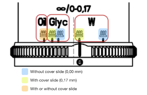
Figure 2: Correction collar of an older Zeiss multi-immersion objektive 40x/0,9 (RMS). This type allows to use different immersion media: Oil, glycerin and water. Water samples always require immersion with a drop of water, however. These multi-immersion objectives are no longer produced. This older series is a recent predecessors of modern high-performance optics, like Zeiss "LCI Plan-Neofluar" und "C-Apochromate W" series.
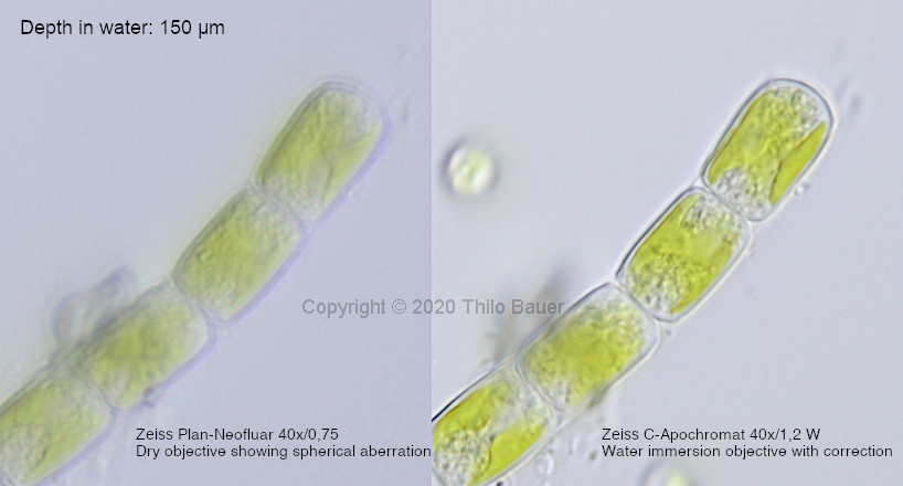
I would like to add two images for comparison and to demonstrate the effect of spherical aberration. Two different objectives were compared to take photomicrographs of the same alga under same conditions. Microscopic slide was prepared of 50 µl sample volume. The alga was located at the bottom of the sample slide preparation and viewed through a water column of 150 µm.
Figure 3: Green alga viewed through thick water column of about 150 µm. Left: Image of a superb dry objective of type Zeiss Plan-Neofluar 40x/0,75. The effect of spherical aberration is clearly seen: The water column introduced loss of contrast, chromatic errors and loss of definition of the focal plane. Right: Same alga viewed with a Zeiss water immersion objective of comparable magnification: C-Apochromat 40x/1,2 W Corr. The water immersion objective easily manages viewing through water column of that size and provides a clear and sharp image.
Summary
Comparison of different dry objectives or oil immersion objectives depends on many factors, when viewing water samples. The image quality suffers from the water sample. Therefore, standard objective types do not yield best or comparable results. Spherical aberration is introduced by light passing different refractive indices of the glass, air or immersion medium, cover slide and water solution. Selection of cover slides with different thickness and also varying height of a water column within the sample preparation lead to large differences in quality of the microscopic image. Hence, one cannot compare dry or oil immersion objectives used for this purpose. Also these objective types are limited to a small range of water column height, short below the cover slide. Careful preparation of samples, like use of precise sample volumes and selected cover slides, will help to improve reproducibility of the image quality. These methods do not overcome spherical aberration, however. If the goal is the best possible contrast, color and resolution of the image, water immersion objectives are the first choice.
Highly diffractive media for observation of cells in combination with high-aperture oil immersion objectives are a special use case for microscopic super-resolution techniques, like TIRF. Manufacturers deliver specialized objective designs for this purpose with numerical aperture of NA>1.4. Such objectives are not designed for observation of water samples or general purpose, however.
Literatur
Vermeulen, K. et al., 2006. Optical trap stiffness in the presence and absence of spherical aberrations. Applied Optics 45(8):1812-9.
S. Bradbury, 2014. Evolution of the Microscope. Elsevier.
Zeiss Operating Manual: Use of the C-Apochromat 40x/1,2 W Corr, publication date unknown.