
 Introduction
Suberin is a polymer found in the cuticula of plant cells. Suberin can be stained with Fluorol Yellow 088 dissolved in polyethylene glycol (PEG) and glycerol. The name of suberin is derived from the corak oat, Quercus suber, which us used to produce cork.
Staining of salvia leaves
A hand-section of a leaf of Salvia officinalis was cut with a razor blade. 5 µl Fluorol Yellow 088 solution were stirred with a water drop. The section was then transferred into this solution on a specimen slide. The preparation was and cover slide. Duration of the staining can be controlled during microscopic observation. The fluorochrome stains suberin fairly quick. A larger section, however, may take a while until it is completely stained. The highly refractive PEG requires some time to completely dissolve in the preparation.
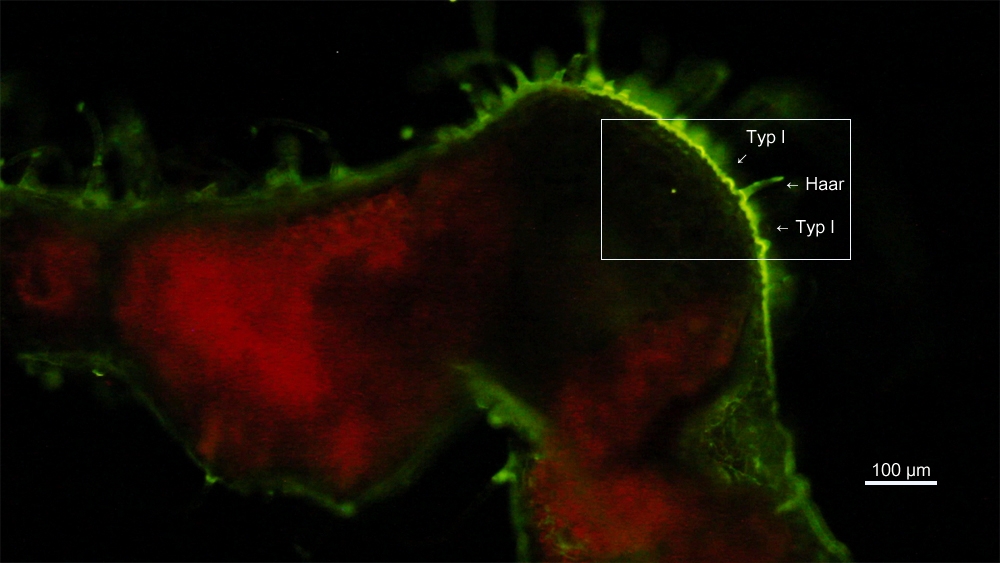
Special literature describes excitation of Fluorol yellow 088 using ultra-violet light (365 nm). This cannot be recommended, however. Absorption and emission spectra of this fluorochrome clearly indicate, it shall be excited using blue light (470 nm) to match its absorption maximum and get the most energy transferred into fluorescence emission. Therefore, fluorescence images were obtained using a blue LED excitation (470 nm). Emission color of Fluorol Yellow 088 is green and best viewed with a long-pass emission filter (λmin=510 nm). Auto-fluorescence of chlorophyll will yield leaf chloroplasts contrasted in red colors. Compared to UV excitation (not shown), excitation with blue light clearly yields brighter fluorescence of Fluorol Yellow 088.

Figure 1: Brightfield image of a hand-section of a leaf of Salvia officinalis taken with an objective of magnification 10x. The surface of the older leaves look somewhat gray or silver. This leaf also showed certain injuries. There are two different types of glandular trichomes (type I and II).
Figure 2: Same image section observed with fluorescence microscopy and excitation at wavelength 470 nm (blue).
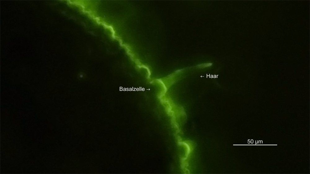 Figure 3: Magnified view of the hand-section near the vein. A microscope objective with magnification 40x was used for this image of the epithelium cells. Suberin is stained well in green color by Fluorol Yellow 088. Suberin is embedded in the cuticle of the epithelium cells (the outer half shell) and glandular trichomes.
Literature
Serrato-Valenti, G., et al. "Structural and histochemical investigation of the glandular trichomes of Salvia aurea L. leaves, and chemical analysis of the essential oil." Annals of Botany 79.3 (1997): 329-336.
Image data
Zeiss AxioLab.A1, N-Achroplan 10x, N-Achroplan 40x, blue excitation (LED 470, nm), Zeiss filter cube 09, Canon EOS 60D.