 Introduction
Epi-fluorescence techniques have been shown to support determination of desmid species using a fluorescence microscope. With auto-fluorescence Netrium species can easily be distinguished from each other from demonstrating the shape of the chloroplasts. The 3D structure of other desmids, like Chlosterium or Micrasterias can be shown well as well. These structures typically cannot be seen using conventional transmission light microscopy.
Experiment
It is not necessary to own any expensive confocal (fluorescence) microscope. A common wide-field fluorescence microscope is just enough to demonstrate the three-dimensional structure of the chloroplasts of any desmids. Fluorescence excitation uses blue light at 470 nm wavelength. With this excitation the chloroplasts of any algae will yield bright red fluorescence using a long-pass fluorescence filter cube. To show the 3D structure water immersion objectives are preferred. This type of objective provides a thin optical cut through the plastids. With focus-stacking the spatial details of the plastids can be well demonstrated.
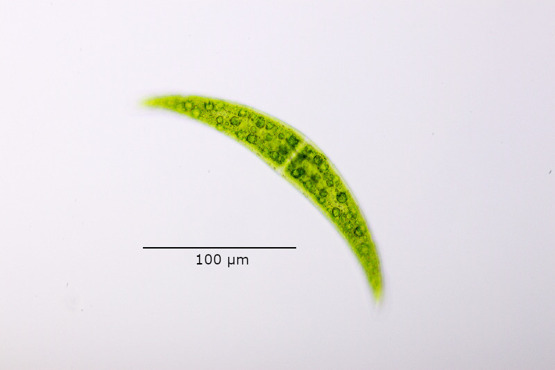
Figure 1: Closterium viewed in a bright field microscope.
Figure 2: This composite shows the same Closterium individual with bright field and different focal planes using auto-fluorescence (470 nm).
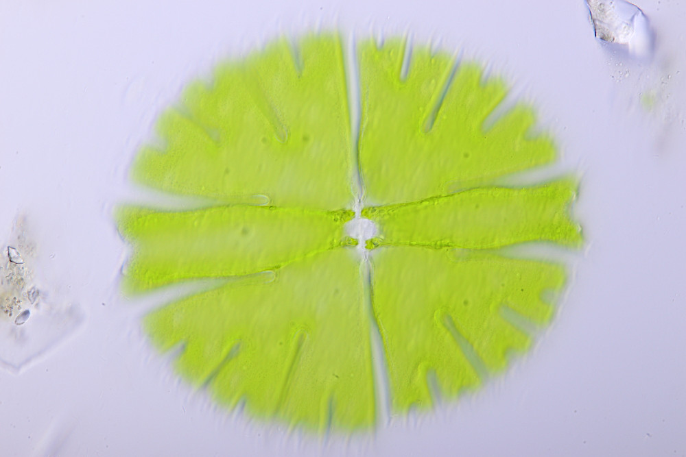
Figure 3: Bright field image of Micrasterias rotata using a Zeiss C-Apochromat 40x/1,2 W korr. The objective with large Aperture of NA=1.2 provides high resolution, but low depth of sharpness.
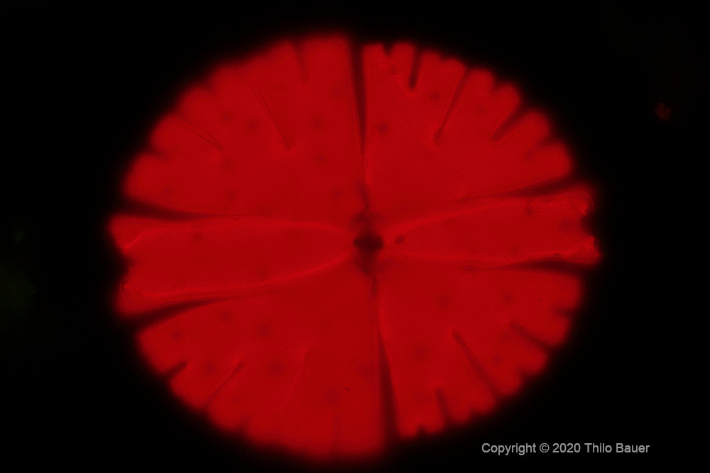
Figure 4: Single fluorescence image of the chloroplast of Micrasterias rotata. A single fluorescence image also provides a thin layer of sharp image only with any high resolution microscope objective.
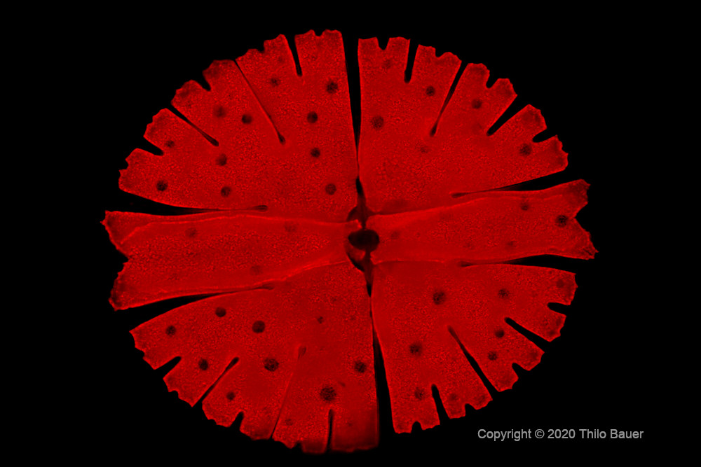
Figure 5: Focus stack of Micrasterias rotata reveals the 3D structure of its chloroplast. Composed of 12 single frames of different focal planes the resulting composite image provides a large depth of sharpness and spatial resolution at best possible optical resolution.
Literature
- Klein, F., 2017. Systematik und Revision der Gattung Netrium auf der Basis morphologischer und molekularer Merkmale unter besonderer Berücksichtigung von Kreuzungsexperimenten und Chromosomenzahlen / Polyploidisierung. Masterarbeit, Universität Köln, im Januar 2017.


