 Introduction
DiOC6(3) is a well-known fluorochrome of the carbocyanine group of stains to selectively stain mitochondria. DiOC6(3) will also stain the membranes of the endoplasmic reticulum (ER) at higher concentration.
The ciliate Frontonia leucas
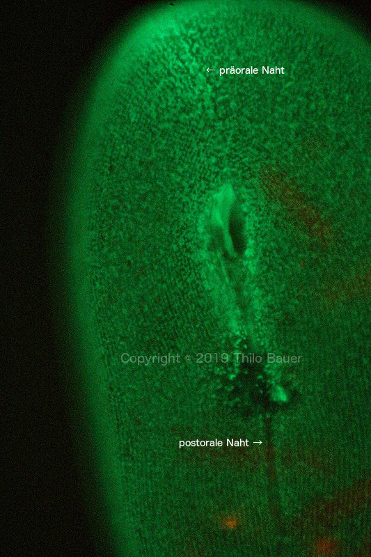
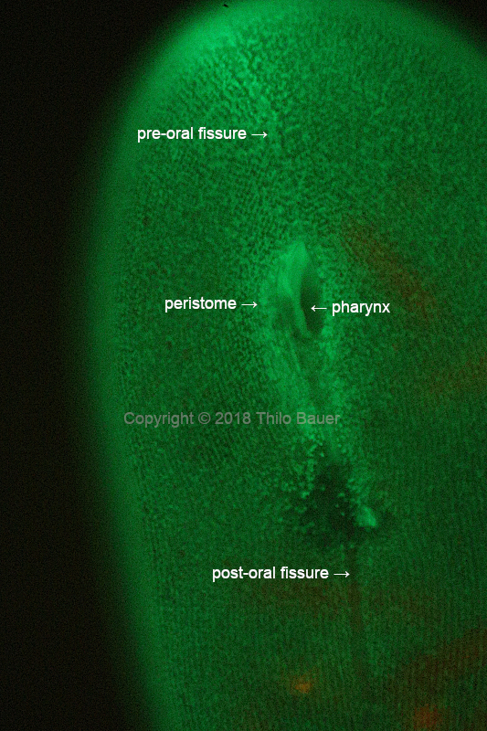
Frontonia leucas is one of the larger ciliate species (phylum Ciliophora). Frontonia leucas reaches a size of up to 600 µm and more. Because of their relatively large size, individuals are visible using a magnifying glass and, looking carefully closer, also to the naked eye. Hence the individual ciliate cell presented here could not be completely viewed using a 40x microscope objective. With fluorescence staining of DiOC6(3) cell viability is well maintained and the single-celled ciliates can be studied using fluorescent life-cell imaging. The latin word "reticulum" translates into the English word "net". With Frontonia the endoplasmic reticulum appears in a very regular pattern of a three-dimensional net of membranes below cell pellicle and filling the whole cell. Close to the pellicle the ER follows the pattern of the ciliary rows. Thousands of cilia are Frontonia's feet to move. In the image presented a pre-oral and post-oral fissure can be clearly seen starting from the large peristome. The fissure is a pattern of ciliary rows endings at the ventral surface of the cell.
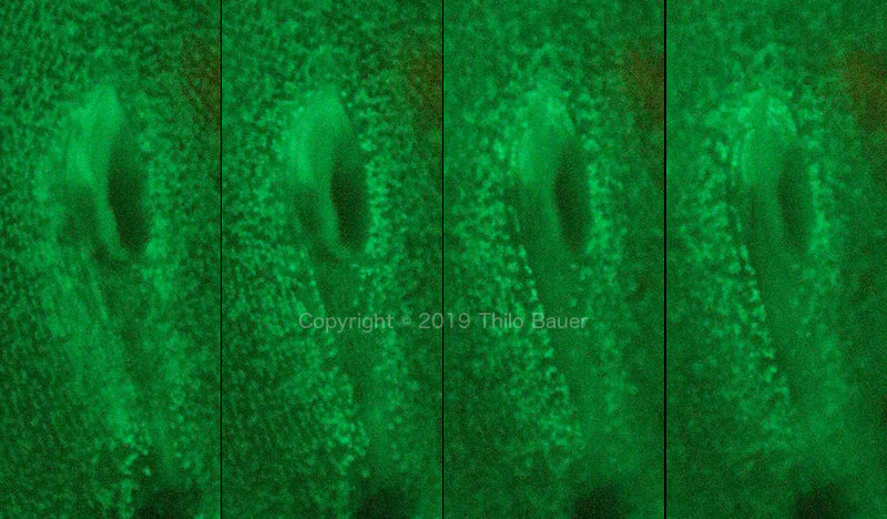
The second image shows details of the peristome when viewed from top of the pellicle to the inner opening. Different depth of the focal planes demonstrate the 3D structure of the pharynx. The endoplasmic reticulum is not a fixed cell organelle, but is constantly changing. This time series was recorded with a conventional digital single-reflex camera. Slight changes of the ER are clearly visible. Also the time series shows slight changes and movement of the pharynx itself.
Image data
Zeiss Axio Lab.A1, Zeiss multi-immersion objektive 40x/0,9 Ph3, blue LED fluorescence excitation (470 nm), Zeiss filter cube No. 09, Canon EOS 77D.


