
 Characteristics
Euplotes daidaleos (Diller & Kounaris, 1966) is one of the large species of the genus Euplotes. Euplotes are a distinct group of unicellular organisms in the group (subclass) of the hypotrichs within the ciliates. Ciliates is a historic name of the enclosing phylum, but still used for these unicellular eucaryotes, that carry cilia used for movement and ingestion. Nowadays the phylum of the ciliates is correctly named as Ciliophora. Genus Euplotes has a characteristic morphology, that makes it easy to determine the genus. Euplotes has a characteristic peristome and a specific pattern of their cirri, which can be observed well in brightfield microscopy. To determine individual species one shall have a look at the size of the cells and morphology, especially the pattern of buccal cirri, transversal cirri and caudal cirri.
The atlas of the ciliates (Foissner et al. 1991) mentions Euplotes daidaleos as a possibility of confusion only and gives no detailed description. However, the species is not really rare to find. From my own experience Euplotes daidaleos is found often in ephemeral waters, like puddles, ponds and also the muddy littoral zone of pools. The individual shown here was found in a pond close to Hürth (close to Cologne, Germany). I described this species earlier in one of my publications demonstrating the success of fluorescent double staining with Hoechst 33342 and acridine orange (Bauer, 2019). The individuals shown here were found in a pool close to the Dörnberg hill (Kassel, Germany). The double staining method is well reproducible and is used to determine ciliate species in general.
E. daidaleos can be separated well within the genus, because individuals bear zoochlorellae (algae). Zoochlorellae are well distinguished from digested algae or diatoms eventually found in phagosomes. See also enlarged view of a chlorella in Fig. 1 (lower right corner). The number of zoochlorelles may vary from a dense population to very few indiviual algae cells located in the corpus of E. daidaleos. If individuals my carry few algae cells only, there is a certain danger that the species E. daidaleos may be falsely determined as a different Euplotes species. E. daidaleos measures about 90 µm in length and 60 µm in width. E. daidaleos also carries eight dorsal ridges with fey cilia. These ridges and smaller cilia can be detected in brightfield imaging at large mignification. More obvious, however, are groups of organelles surrounding the kinetosomes (basal bodies) of the dorsal cilia. To observe these organelles, one shall focus carefully at the dorsal surface. Dorsal organelles are arranged in rings around the kinetosomes. These rings are composed of small acidic vacuoles and mitochondria. Double staining using Hoechst 33342 and acridine orange yields good results to determine ciliate species in fluorescence (Bauer, 2019). Dorsal vesicles (dV) are acidic organelles which are stained with acridine orange and provide green to yellow or red fluorescence (see also Fig. 3). Mitochondria will not be stained by acridine orange, but are visible in brightfield microscope at higher magnification from their large diffractive index (see images from Bauer, 2019). Euplotes species have a characteristic macronucleus, formed like the character "C" or the digit "3". In my images the macronucleus (Ma) is well seen from fluorescence staining with Hoechst 33342. Chromatin of the macronucleus often appears granulated, like a network or vesicular. The granluated structure of the macronucleus of Euplotes daidaleos is well seen from the specific staining of Hoechst 33342 in my images. Close to the macronucleus a single micronucleus is found in Euplotes species. One pulsating vacuole is located in the rear third of the corpus (figure 1).
Legend
AZM: Peristome, adoral zone of membranelles
Ma: Macronucleus
Mi: Micronucleus
Ph: Phagosome
Chl: Zoochlorella
cV: Contraktile vakuole
dV: Dorsal vesicles, surrounding dorsal kinetosomes
CC: Caudal cirri
TC: Transversal cirri
Figure 1: Euplotes daidaleos, dorsal view. Brightfield photomicrograph taken with a water immersion objective and optimized oblique illumination.

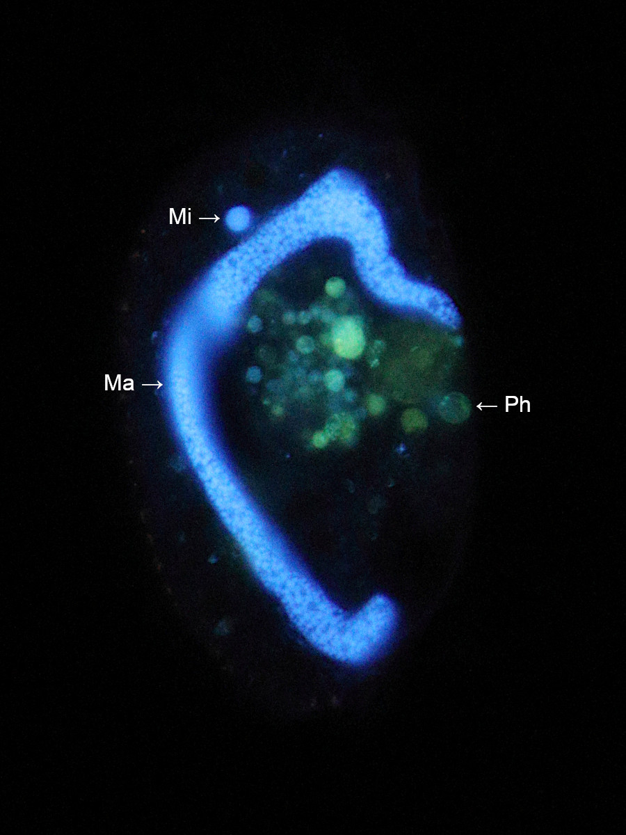
Figure 2: Same individual as shown in figure 1. Photomicrograph taken in epi-fluorescence illumination with double staining of Hoechst 33342 and acridine orange. UV excitation (385 nm). The granulated macronucleus (blue) is well visible together with the small micronucleus. Bakterial DNA digested in a few phagosomes appear in blue-green colors.
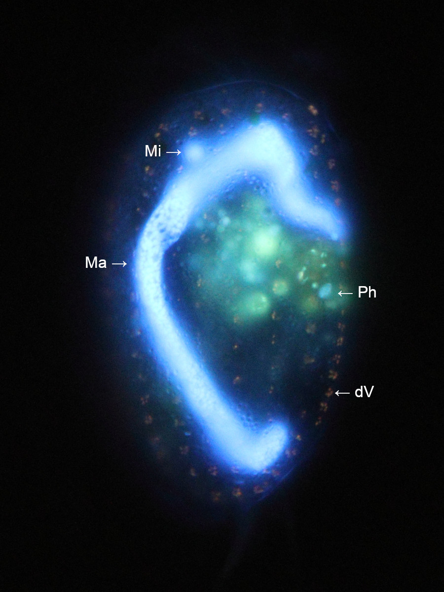
Figure 3: From longer exposed and contrasted fluorescence images, taken in the focal plane of the dorsal pellicle, acridine orange stains acidic vesicles surrounding dorsal kinetosomes. Dorsal ridges and cilia are hard to detect from brightfield observation, but the organelles surrounding the kinetosomes (basal bodies) are visible at large magnification.
Imaging details
Zeiss Axio Lab.A1, water-immersion objektive Zeiss C-Apochromat 40x/1.2 W korr with condenser 1,25. Brightfield imaging with optimized oblique illumination. Fluorescence: Triple-band filter cube F66-412, UV excitation at 385 nm. Camera: Canon EOS 77D.
Literature
- Diller, W. F., and Kounaris, D., 1966. "Description of a zoochlorella-bearing form of Euplotes, E. daidaleos n. sp. (Ciliophora Hypotrichida)." The Biological Bulletin 131.3 (1966): 437-445.
- Foissner, W. et al., 1991. Taxonomische und ökologische Revision der Ciliaten des Saprobiensystems, Informationsberichte des Bayer. Landesamts f. Wasserwirtschaft.
- Bauer T., 2019. Fluoreszenz-Doppelfärbung mit Hoechst 33342 und Acridinorange zur Bestimmung der Arten von Ciliaten (Phylum: Ciliophora). Mikroskopie. 2019; 1: 2-19.