
Introduction
Stylonychia mytilus denotes a complex of species within the unicellular group of ciliates (Phylum: Cilophora). Stylonychia mytilus consists of at least two similar, but genetically different species: S. mytilus und S. lemnae (Berger, 1999).
Determination & fluorescence staining
Typical for Hypotricha are cirri. Cirri are bundles of fine cilia. It appears, as if hypotrichs "crawl by feet", which is the reason why they sometimes are called "Lauftierchen" (in German). Individual cilia at the ends of the cirri can be seen with medium to large magnification using a light microscope at magnification >40x. Determination of species within the family of the hypotrichs is hindered by large rates of cell division. During cell division the pattern of cirri and cilia changes. On the other side the pattern of cirri is important for species determination. Especially in fresh samples or cultures cell division can be raised significantly. Hypotrichs can be watched at low magnification even in an stereo microscope. Here they tend to show jerky movement, jumping back and forth.
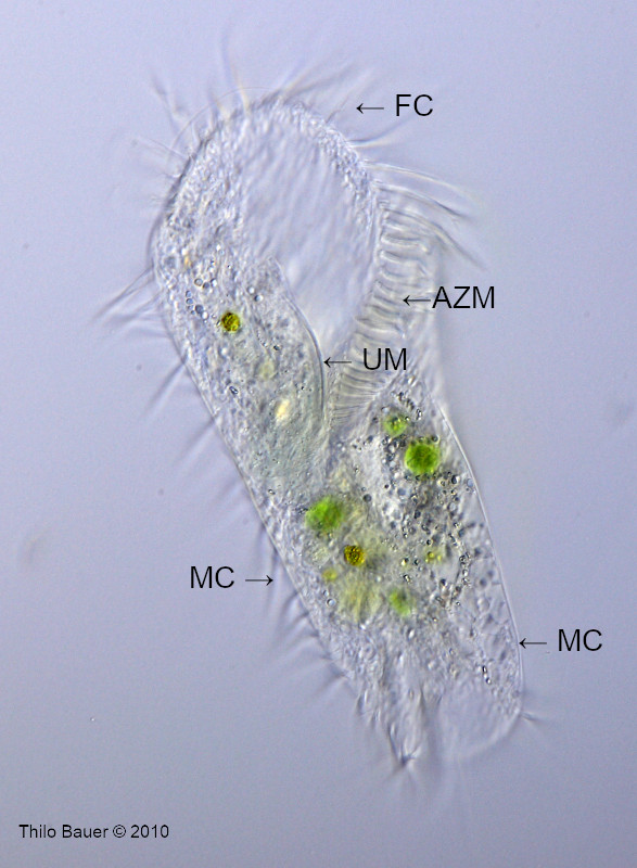
Stylonychia mytilus is one of a few hypotrichs, that can be easily determined. Stylonychia mytilus is a ciliate of striking size of 100-400 µm. Looking onto the ventral side, a broad head with peristome, undulating membrane and adoral zone of membranelles (AZM) is protruding towards the right side (Fig. 1). Flanks are parallel to slightly cone-shaped towards the tail. At low magnification in stereo microscope Stylonychia mytilus looks somewhat "rectangular with a hole", that is the peristom at head. To properly determine and demonstrate this species, microscopic squash preparation shall be avoided. Instead, free swimming individuals shall be observed to demonstrate their natural shape.
The special patterns of front cirri, ventral cirri, transversal cirri and caudal cirri, where they exist, are characters of the familiy Hypotricha. Berger (1999) compares different sketches of Stylonychia mytilus as depicted by different authors. Subtle differences of the pattern of the cirri will confuse the beginner also in case of oxytrichs, like Stylonychia mytilus. Also important are marginal rows of cilia in case of Stylonychia mytilus.
Ciliates usually have two different kinds of nuclei, macronuclei and micronuclei. These can hardly be distinguished from other organells, esp. if ciliates are in late cell cycle and well-fed. Few has been written about micronuclei of many species, because they are often hard to find. The technique of double fluorescence staining supports well determination of nuclei in living ciliates.
Figure 2 shows details about the pattern of dorsal cilia (DC), that appear like small spikes arranged in six rows. These dorsal cilia are character to distinguish between S. mytilus and S. lemnae (Berger, 1999). Photomicrography skills are advanced to demonstrate these cilia, however. It is important to note that the number of dorsal cilia in rows 3 and 4 is lower for S. lemnae, when compared to the larger S. mytilus.
AZM: Adorale Membranellenzone
CC: Caudalcirren
cV: kontraktile Vakuole
DC: Dorsale Cilien
FC: Frontalcirren
MC: Marginalcirren (=randliche Cirren)
Ma: Macronucleus
Mi: Micronucleus
Ph: Phagosom
UM: Undulierende Membran
Figure 1: Sytlonychia mytilus (lemnae?) free swimming individual viewed from ventral side.
Figure 2: Stylonychia mytilus (lemnae?) viewed from ventral side. Here the focal plane was shifted to the nuclei. As the individual is oriented in 2D space, view is directed through the cell to the back, where dorsal cilia (DC) can be seen. Short marginal cilia (MC) at the left and right side form row number #1 and #6 of the dorsal cilia. Three large caudal cilia are important characters of some oxytrichids, like Stylonychia mytilus.
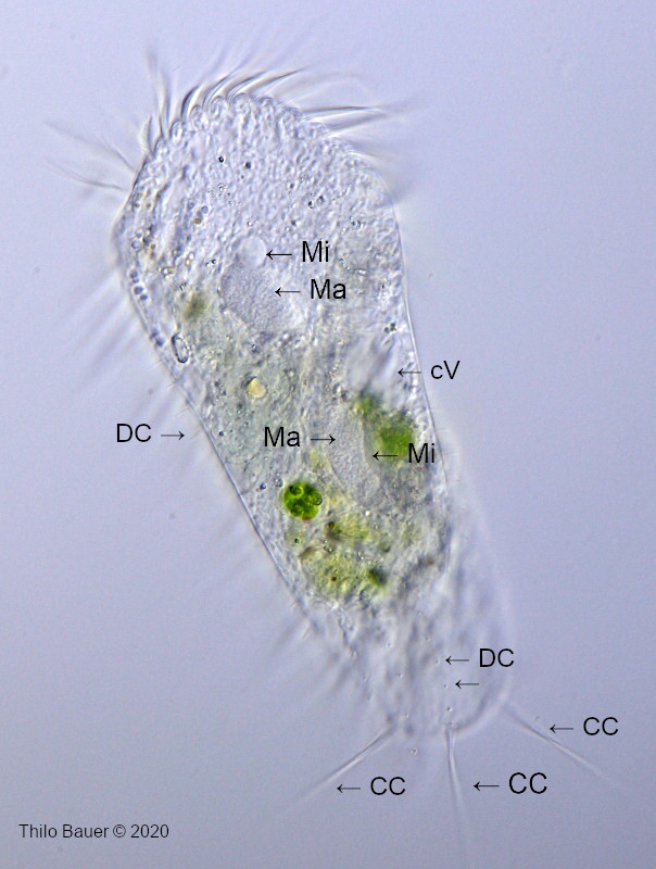
Figure 3: Stylonychia mytilus (lemnae?) demonstrated in fluorescent double staining and UV illumination. The two forms of nuclei can be well determined.

Observational data
Pictures taken with Zeiss Axio Lab.A1 FL-LED, Zeiss C-Apochromat 40x/1,2 W, condensor 1,2 and water immersion. Bright field images taken in optimized oblique illumination. Fluorescence image: Double staining with Ho342 and acridine orange, triple-band filter F66-412, UV excitation (385 nm). Camera: Canon EOS 77D.
Literature
Berger, H., 1999. Monograph of the Oxytrichidae. Kluwer Academic Publishers.
Foissner, W. et al., 1991. Taxonomische und ökologische Revision der Ciliaten des Saprobiensystems.